If a doctor has suggested you might have pulmonary hypertension, or if you are already partway through testing, the list of tests can feel overwhelming. Terms like echocardiogram, right heart catheterization, and vasoreactivity testing may sound intimidating before you understand what they involve. Each test has a clear purpose, though, and knowing that purpose can make the whole process feel more manageable.
This article walks through the most common tests used to screen for, diagnose, and classify pulmonary hypertension, explains what each one measures, and describes what typically happens after a diagnosis is confirmed. Talk with your care team about what approach makes sense for your specific situation.
What Is Pulmonary Hypertension, and Why Is It Hard to Spot Early?
Pulmonary hypertension (PH) is a condition in which the blood pressure inside the arteries that carry blood from the heart to the lungs becomes abnormally elevated. Over time, the increased resistance forces the right side of the heart to work harder, which can eventually lead to right heart strain or failure if left unmanaged.
One reason PH can be difficult to catch early is that its most common symptoms, shortness of breath during activity, fatigue, and dizziness, are shared by many other, more common conditions. People sometimes attribute these feelings to being out of shape, aging, or stress. As a result, there is often a significant gap between when symptoms first appear and when a correct diagnosis is made.
That delay is exactly why a structured diagnostic process matters. The tests described below are designed to build on one another: starting with lower-risk screening tools and progressing toward more definitive procedures when needed.
The Echocardiogram: The First Major Clue
For most people, testing begins with an echocardiogram, a non-invasive ultrasound of the heart. During this test, a technician uses a handheld device called a transducer to send sound waves through the chest wall. These waves bounce off heart structures and create real-time images of the heart's chambers, valves, and blood flow patterns.
An echocardiogram can do several important things in the context of pulmonary hypertension:
- Estimate pulmonary artery pressure. By measuring the speed of blood flowing backward through the tricuspid valve (a phenomenon called tricuspid regurgitation), the echo technician and reading cardiologist can estimate the pressure in the pulmonary arteries. This estimate is not a direct measurement, but it provides a meaningful signal that something may be wrong.
- Assess right heart size and function. An enlarged or poorly functioning right ventricle can be a sign that the heart has been working against elevated pressure for some time.
- Look for structural causes. Certain heart defects, such as holes between heart chambers, can cause elevated pulmonary pressures. An echocardiogram can often identify these.
It is important to understand that an echocardiogram screens for pulmonary hypertension rather than definitively diagnosing it. The pressure estimates it provides can be influenced by technical factors and individual anatomy. If the echo raises concern, further testing is almost always needed.
Other Imaging and Functional Tests
Before or alongside a referral for more invasive testing, clinicians often order several additional studies to build a complete picture. These may include:
- Chest X-ray. A plain chest X-ray can show enlargement of the pulmonary arteries or the right heart and may reveal underlying lung conditions contributing to elevated pressures.
- CT pulmonary angiography (CTPA) or high-resolution CT scan. These detailed scans help identify chronic blood clots in the lung arteries (a condition called chronic thromboembolic disease) or underlying lung diseases such as interstitial lung disease, which are important causes of pulmonary hypertension.
- Ventilation-perfusion (V/Q) scan. This nuclear medicine test looks at how air and blood flow through the lungs and is particularly useful for detecting mismatches that suggest chronic clots.
- Pulmonary function tests (PFTs). Breathing tests measure lung capacity and airflow, helping to identify or rule out obstructive and restrictive lung diseases.
- Six-minute walk test. This simple exercise assessment measures how far a person can walk in six minutes and provides a snapshot of functional capacity and how well the heart and lungs are working together under mild exertion.
- Blood tests. A panel of blood work, including markers of heart stress, thyroid function, connective tissue disease, and liver function, helps identify underlying causes and assess overall health.
Together, these tests help clinicians classify the type of pulmonary hypertension, which matters because different types require different treatments.
Right Heart Catheterization: The Gold Standard
When clinical suspicion for pulmonary hypertension remains after non-invasive testing, the next step is typically a right heart catheterization (RHC). This is the only test that can directly and accurately measure the pressures inside the pulmonary arteries, and it is considered the definitive diagnostic procedure for PH.
Here is what to expect:
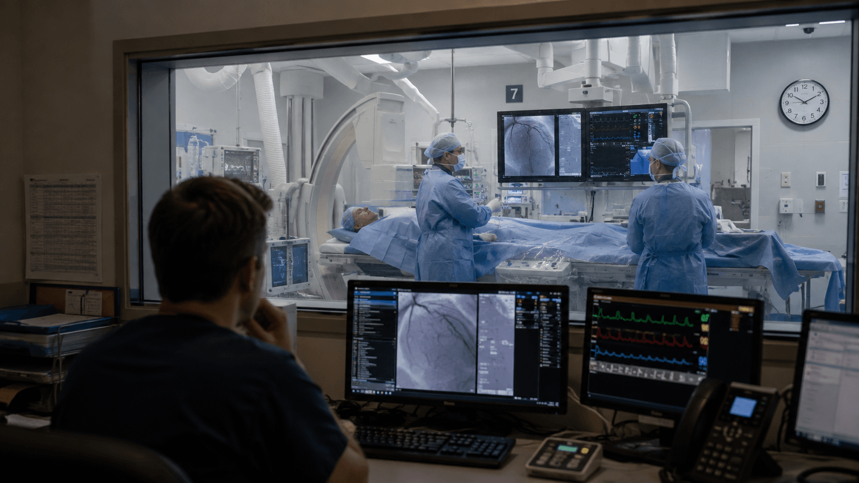
The procedure is usually performed in a cardiac catheterization laboratory (cath lab) by an interventional cardiologist or pulmonologist. You will likely receive a mild sedative to help you relax, though you typically remain awake. A thin, flexible tube called a catheter is inserted, most often through a vein in the neck, groin, or wrist, and carefully guided through the right side of the heart and into the pulmonary arteries.
As the catheter travels through each chamber, it measures pressures directly. Clinicians record several key values, including:
- Mean pulmonary artery pressure (mPAP). The average pressure in the pulmonary arteries. A value above a specific threshold, measured at rest, is one of the criteria used to confirm a diagnosis of pulmonary hypertension.
- Pulmonary capillary wedge pressure (PCWP). This measurement helps distinguish between PH caused by left heart disease (where the wedge pressure is elevated) and PH arising from the pulmonary arteries themselves (where it is typically normal).
- Cardiac output. How much blood the heart pumps per minute. This, combined with the pressure readings, helps calculate pulmonary vascular resistance, a measure of how hard the heart must work to push blood through the lungs.
The procedure usually takes less than an hour, and most people go home the same day. It is generally well-tolerated, though as with any invasive procedure, your care team will discuss the small risks involved before you consent.
Vasoreactivity Testing: A Key Step for Some Patients
For a subset of patients, particularly those with a specific subtype called pulmonary arterial hypertension (PAH), doctors may perform vasoreactivity testing during the right heart catheterization. During this add-on test, a short-acting medication (often inhaled nitric oxide or another agent) is given to relax the pulmonary blood vessels, and pressures are re-measured.
A meaningful drop in pulmonary artery pressure in response to this medication, called a positive vasoreactivity response, identifies patients whose condition may respond well to high-dose calcium channel blockers, a class of medication that is generally not effective for PH unless this response is present. This distinction can shape the treatment plan.
What Comes Next After Diagnosis
Receiving a diagnosis of pulmonary hypertension is understandably a lot to absorb. The path forward depends heavily on what type of PH you have, its severity, your overall health, and the underlying cause.
A few general things to know:
- Classification matters for treatment. Pulmonary hypertension is grouped into several categories based on its cause. Treatment that is highly effective for one type may not be appropriate for another, and could even be harmful. This is why the thorough diagnostic workup described above matters before any medications are prescribed.
- Specialist care is strongly recommended. PH is a complex condition, and guidelines from major cardiology and pulmonology societies recommend that patients be evaluated at centers with expertise in pulmonary hypertension. Specialists in this field stay current with rapidly evolving treatment options.
- Treatment options have expanded significantly. Depending on the type and severity of PH, management may include medications that work on several different biological pathways to relax and open the pulmonary arteries, reduce pressure, and protect the right heart. Some patients are also candidates for specific surgical or interventional procedures.
- Lifestyle and monitoring play an ongoing role. Regular follow-up visits, repeat six-minute walk tests, and periodic echocardiograms or catheterizations help your care team track how your condition is responding over time and adjust your plan as needed.
A note on advocacy: If you feel your symptoms have not been fully explained, or if you are waiting a long time for answers, it is entirely appropriate to ask for a referral to a pulmonary hypertension specialist or to seek a second opinion. Early, accurate diagnosis gives you the widest range of options.
The Bottom Line
Diagnosing pulmonary hypertension is a step-by-step process that moves from non-invasive screening with an echocardiogram through a series of targeted tests to the definitive measurement provided by right heart catheterization. Each step adds information that helps clinicians identify whether you have PH, and also what type it is and why, questions that directly shape your treatment.
If you or someone you love is going through this process, know that asking questions, understanding what each test is looking for, and building a relationship with a knowledgeable care team are all ways to stay engaged with your own health. Speak with your clinician about any concerns, and do not hesitate to ask what the results of each test mean for the next step in your care.